Figure 1.
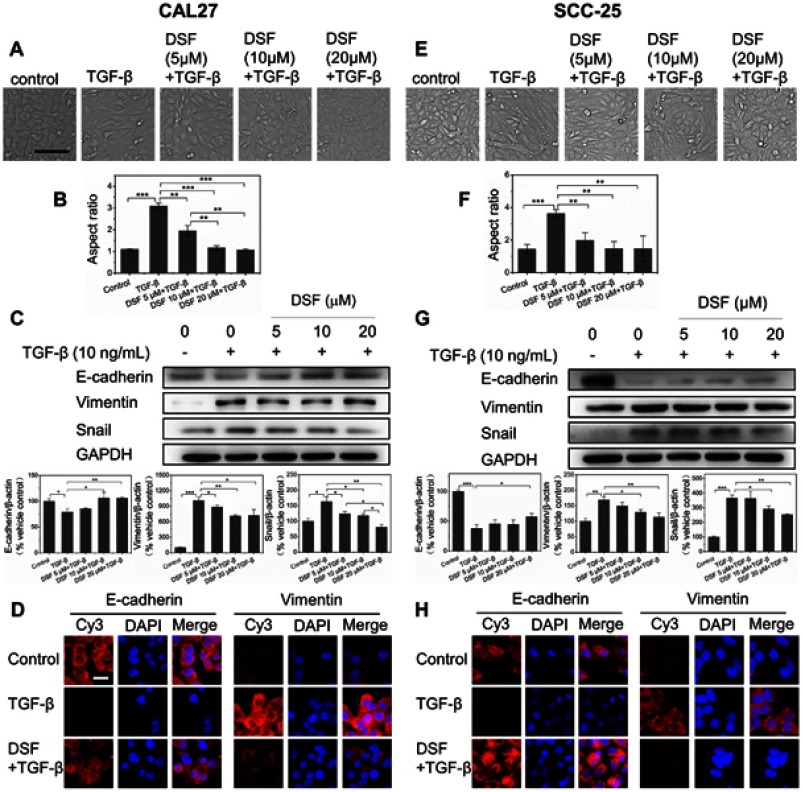
Dsf inhibited TGFβ1-induced EMT in OSCC cells.
Notes: EMT in OSCC cells was induced by TGFβ1 stimulation for 6 days. Dsf was added to cells 24 hours before TGFβ1 induction to inhibit EMT. (A, E) Morphological changes in OSCC Smad4-mutated CAL27 and Smad4 wild-type SCC25 cells after stimulation with TGFβ1 (10 ng/mL) alone or combined with Dsf (5, 10, 20 μM) were observed under light microscopy. Bar 100 μm. (B, F) Aspect ratios of cells were quantified. Values represent mean ± SEM. (C, G) Western blot was performed to detect the expression of E-cadherin, vimentin and Snail in cells treated with TGFβ1 (10 ng/mL) alone or combined with Dsf (5, 10, 20 μM); quantitative results of Western blot from three independent experiments shown as mean ± SEM. (D, H) Expression of E-cadherin and vimentin examined through immunofluorescence assay by fluorescence microscopy in cells treated with TGFβ1 (10 ng/mL) alone or combined with Dsf (20 μM). *P<0.05; **P<0.01; ***P<0.001; bar 20 μm.
Abbreviations: Dsf, disulfiram; OSCC, oral squamous cell carcinoma; EMT, epithelial–mesenchymal transition.

