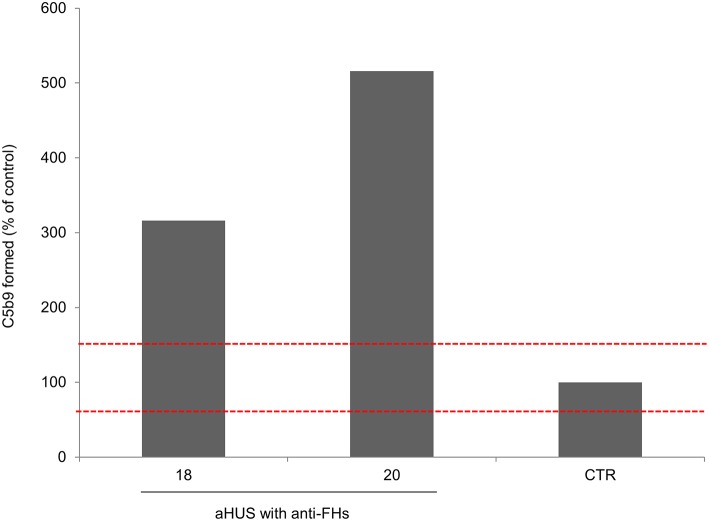
Figure 4.
Complement activation on endothelial cells. Endothelial surface area covered by C5b-9 staining after incubation of ADP-activated HMEC-1 with serum from aHUS patients with anti-FHs studied at remission (patients 18 and 20). For each sample, values were expressed as the percentage of C5b-9 deposits induced by a pool of sera from 10 healthy controls run in parallel (reference 100%). The red dashed lines indicated the normal range (60–149%) determined by testing single sera from 35 different healthy controls.

