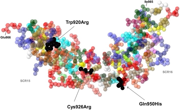
Figure 5.
3D structure by nuclear magnetic resonance of SCR15 and SCR16 of FH (PDB code: 1HFH). Black arrows indicate the FH amino acid residues Trp920, Cys926 (SCR15), and Gln950 (SCR16) (colored in black) which are substituted in three aHUS patients with anti-FHs due to the Trp920Arg, Cys926Arg, and Gln950His heterozygous mutations. Each amino acid is represented with a specific color. The first amino acid of SCR15 (Glu866) and the last amino acid of SCR16 (Ile985) are also indicated. Cysteine residues are shown in yellow color. Created by Jmol, an open-source Java viewer for chemical structures in 3D (http://www.jmol.org).

