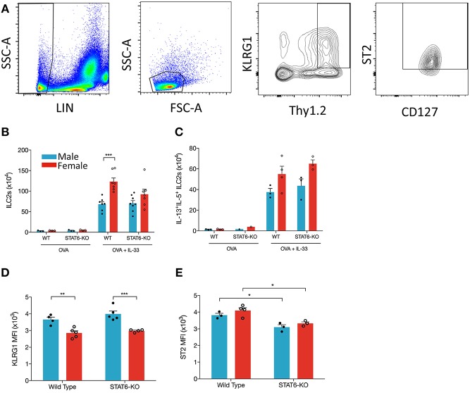
Figure 6.
IL-33 induces ILC2 proliferation more effectively in female mice and is STAT6-independent. Mice were treated as in Figure 1A. Total lung cells were stained as described in the methods. (A) ILC2s were identified as Lin-, small lymphoid-like cells, Thy1.2hiKLRG1+ST2+CD127+ and quantified (B) as absolute cell number. (C) ILC2s expressing both IL-13 and IL-5 were quantified as absolute number. (D) KLRG1 MFI and (E) ST2 MFI of ILC2s were defined. Data in (B) are the combination of two independent experiments (n = 7–8), except for OVA controls (n = 3) and presented as mean + SEM. Date in (C) are from a single experiment (n = 3–4, expect STAT6-KO mice where n = 1). Data in (D) are from a single experiment (n = 4–5). Data in (E) are from a single experiment (n = 3–4). Two-way ANOVA, Tukey's post hoc test. *p ≤ 0.05, **p ≤ 0.01, ***p ≤ 0.001.

