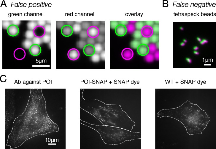
Figure 1.
Image errors can lead to incorrect results. (A) Bleed-through causes a false-positive colocalization result. Green and red beads were mixed and mounted together. There are no beads in these samples/images that are labeled with both green and red dye. With this filter and sample combination, there is significant bleed-through from the green beads into the red channel (see green circles). Pearson’s R for colocalization is 0.67. Since no pixel that contains green fluorophore also contains red fluorophore, there should be no correlation. (B) Channel misregistration causes a false-negative colocalization result. Tetraspeck beads are labeled with four dyes, including dyes imaged in the green and red channels here. Because each bead is labeled with both dyes, there should be complete colocalization between channels, with an expected Pearson’s R of 1. However, the imaging system has introduced significant misregistration between the channels, leading to a Pearson’s R of 0.66. (C) Nonspecific dye binding leads to a false-positive result. A significant level of nonspecific binding of SNAP dye to cells containing no SNAP tag (WT + SNAP dye) looks qualitatively similar to both cells containing a SNAP tag fused to the POI and immunofluorescence against POI. White dotted lines indicate cell outlines.

