Figure 4.
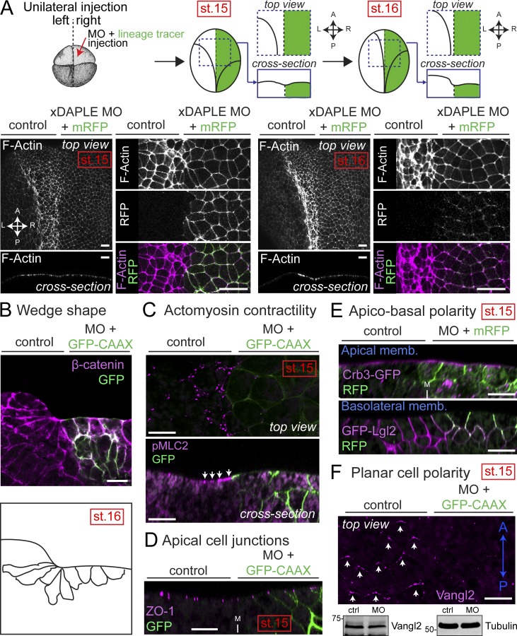
Loss of DAPLE in Xenopus causes apical constriction defects during neurulation. (A) Whole-mount F-actin staining (magenta) of Xenopus embryos unilaterally coinjected with xDAPLE MO and a lineage tracer (mRFP or GFP-CAAX, green) showing enlarged apical surface of DAPLE-depleted neuroepithelial cells compared with uninjected control sides at stages 15 and 16. (B) Transversal cryosection stained for β-catenin (magenta) of the anterior neural plate of an embryo at stage 16 unilaterally coinjected with xDAPLE MO and a lineage tracer (GFP-CAAX, green). Outlines of cell borders are depicted in the bottom to show the lack of wedge shape morphology in the outer layer of neuroepithelial cells depleted of DAPLE. (C–F) Whole-mount pMLC2 (C), ZO-1 (D), GFP (E), and Vangl2 (F) staining (magenta) of Xenopus embryos unilaterally coinjected with xDAPLE MO and a lineage tracer (GFP-CAAX or mRFP; green). In E, embryos were bilaterally injected with Crb3-GFP (top) or GFP-Lgl2 (bottom). In F, staining for the lineage tracer (mRFP) is not shown for clarity, and an immunoblot from dissected neural plates is shown in the bottom. xDAPLE depleted sides show defective staining for actomyosin contractility and PCP markers at stage 15, while markers of apical cell junctions or apicobasal polarity are not changed. All images presented in this figure are representative results of n ≥ 3 experiments. All scale bars represent 25 µm, except those in A, which represent 50 µm.

