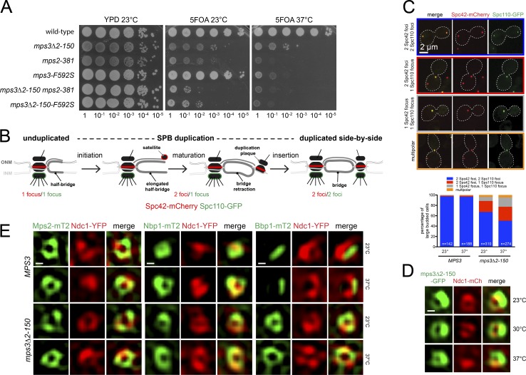
Figure 4.
Role of the Mps3 N terminus in SPB duplication. (A) Serial dilution assay to test growth of wild-type and single and double mutants containing pURA3-MPS3 (SLJ1053) on 5-fluoro-orotic acid at 23°C (3 d) and 37°C (2 d). As a control, cells were also stamped to YPD at 23°C (2 d). Note, mps3Δ2-150, mps3-F592S, and mps2-381 all encode stable proteins (Jaspersen et al., 2006; Gardner et al., 2011). (B) Schematic of SPB duplication pathway from unduplicated to duplicated side-by-side SPBs. Incorporation of Spc110 (green) into the new SPB requires its insertion into the NE, whereas Spc42 (red) localizes to the new SPB early in duplication before insertion. Defects in different steps of SPB duplication can be determined by counting the number of Spc42 and Spc110 foci (Pereira et al., 1999). (C and D) Images showing distinct localization patterns of Spc42-mCherry (red) and Spc110-GFP (green). The percentage of MPS3 (SLJ12982) or mps3Δ2-150 (SLJ12981) cells grown at 23°C or shifted to 37°C for 3 h with each is shown below. (D) SIM of mps3Δ2-150-GFP (green) and Ndc1-mCherry (red) from cells (SLJ12541) grown at 23°C or shifted to 30° or 37°C for 3 h. (E) Localization of Mps2-mT2 (SLJ10472/SLJ13023), Nbp1-mT2 (SLJ8341/SLJ13024), and Bbp2-mT2 (SLJ9231/SLJ13025; green) to SPB toroid by SIM using Ndc1-YFP (red) as a reference, in MPS3 or mps3Δ2-150 grown at 23°C or shifted to 37°C for 3 h. Bars, 100 nm unless indicated otherwise.

