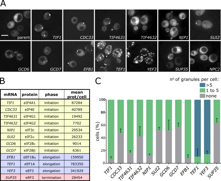
Figure 1.
Translation factor mRNAs localize to cytoplasmic granules in exponentially growing S. cerevisiae. (A) Z-stacked images of strains expressing the labeled MS2-tagged mRNAs and the MS2 coat protein GFP fusion (MS2-CP-GFP). Bar, 4 µm. (B) The tagged mRNAs, proteins they encode, translation phase they function in, and mean protein (prot.) number per cell are listed (from Ho et al., 2018). (C) Chart showing the percentage of cells with one to five, more than five, or no granules per cell. Error bars = +SD (n = 3, >100 cells per repeat).

