Figure 4.
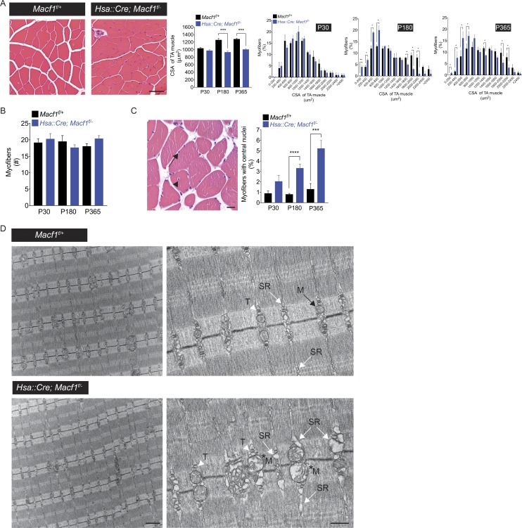
Muscle fibers are atrophic and show aberrations in the structure of the SR and mitochondria in Macf1 mutant mice. (A) Cross-sections of P30, P180, and P365 tibialis anterior muscles were stained with hematoxylin and eosin. The cross-sectional area (CSA) of myofibers is reduced by 30% in Hsa::Cre; Macf1f/− mice at P180 and P365. The mean ± SEM values from three mice (100 myofibers per mouse) are shown (***, P < 0.0005). Bar, 50 µm. (B) The number of myofibers is normal in Macf1 mutant mice. The mean ± SEM values from three mice (measured area: 40,000 µm2) are shown. (C) Macf1 mutant mice exhibit a low level of muscle degeneration/regeneration. 1% of myofibers in control mice and 3–5% of myofibers in Macf1 muscle-conditional mutants have centrally located nuclei (arrow). The mean ± SEM values from three mice (100 myofibers per mouse) are shown (***, P < 0.0005, ****, P < 0.00005). Bar, 10 µm. (D) The organization of sarcomeres, Z-discs, and T-tubule/SR triads (arrowhead) appears normal in the absence of MACF1. In Macf1 mutant mice, the SR (white arrow) is distended, ranging from modestly to severely enlarged, and a subset of mitochondria (*M) shows signs of damage. Bar, 1 µm for the left images and 500 nm for the right images.

