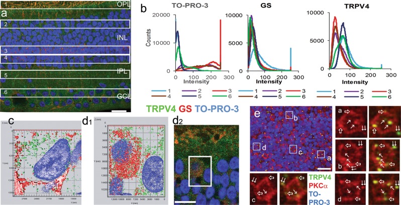
Fig. 2. TRPV4 immunoreactivity in retinal neurons and Mȕller cells.
Monkey retinal slices (a–d) were labeled for TRPV4 (LS-C135), glutamine synthetase (GS) and TO-PRO-3. b shows pixel histograms of TRPV4 immunoreactivity in 6 same sized retinal zones in a (1-the outer plexiform layer, 2-the bipolar cell soma layer (BCL), 3-the Mȕller cell soma layer (MCL), 4-the amacrine soma layer (ACL), 5- the inner plexiform layer and 6-the RGC soma layer (GCL). GS-positive somas are primarily located in Zone 3, where the linear density of TO-PRO-3 labeled nuclei is higher than that in Zone 2 and 4 (ratio: 1.8: 1.2: 1) (a and b). TRPV4 pixel histograms generally fall into two groups, one for those from Zone 1, 5, and 6 and the other for those from Zone 2, 3, and 4 (b). c and d1 are the surface profile of 3D projections of 0.9 μm-thick blocks in the GCL (c) and BCL (d1), and TRPV4 puncta are not fully colocalized with GS. d1 displays the inset of d2. In e, a flat-mount monkey retina was labeled for TRPV4 (LS-C94498, green), PKCα (red), and TO-PRO-3 (blue). The confocal micrograph shows the optical section of the BCL, where TRPV4 puncta are colocalized with PKCα inside the somas (arrow), somatic membrane (open arrow) and dendrites (double arrow) of rod bipolar cells (RBCs). TO-PRO-3 labels nuclei, Scale bars are 20 μm

