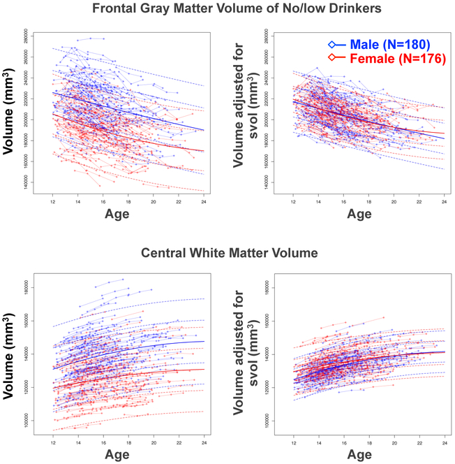
Figure 1.
Left: Plots of 356 adolescents who continued to meet no/low criteria with connected baseline, 1-year, and 2-year follow-up values (boys in blue, girls in red) plotted as a function of their baseline age for frontal gray matter volume (top) and the central white matter sample volume (bottom). The lmer fits with +/− 1 and 2 SD separately computed for boys (blue) and girls (red) are also plotted. Note that the frontal lobe values decrease fairly linearly, whereas the growth in the white matter volume slows over age. For both regions, boys had larger volumes than girls.
Right: The same data as above but adjusted for variation in supratentorial volume (svol), which attenuated sex differences in regional volumes.

