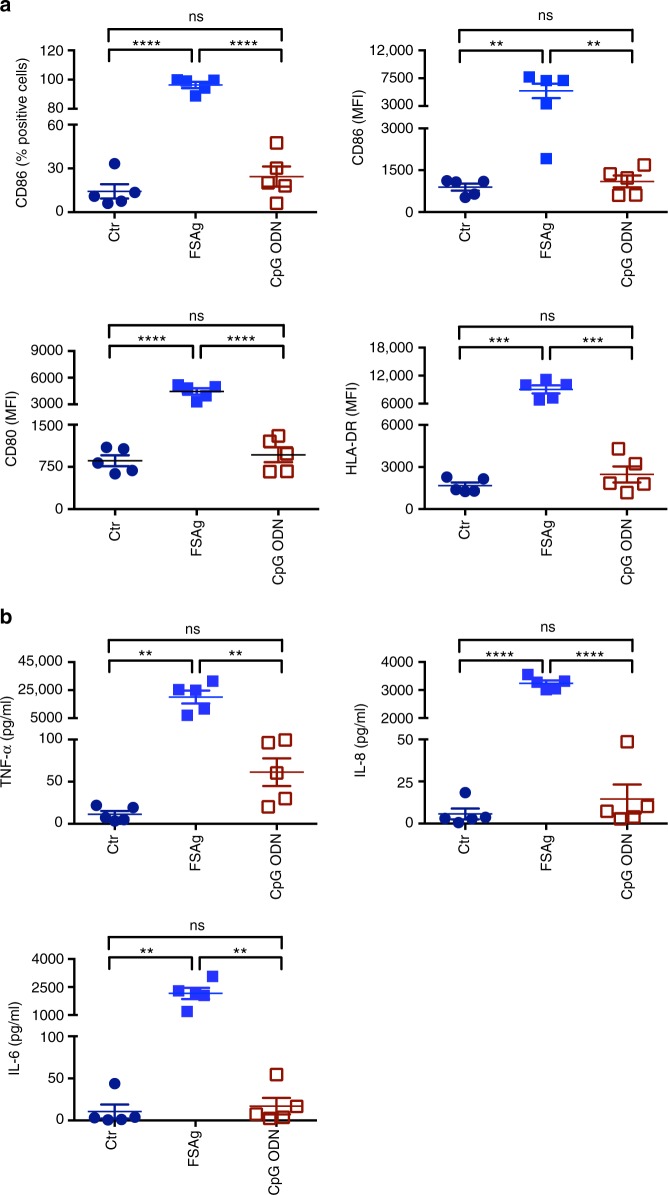
Fig. 4.
Comparison of dendritic cell stimulatory capacity of W. bancrofti sheath antigen and CpG ODN 2006. a Expression of dendritic cell-maturation markers CD86, CD80 and HLA-DR (mean±SEM; % positive cells or median fluorescence intensities, MFI) upon stimulation of cells with microfilarial sheath antigen (FSAg) or CpG ODN. Data are from five independent donors. b Amount of secretion (mean±SEM, n = 5) of dendritic cell cytokines TNF-α, IL-8, and IL-6 (all in pg/ml) under above stimulatory conditions. **P < 0.01; ***P < 0.001; ****P < 0.0001; ns, not significant as analyzed by one-way ANOVA test. Abbreviation: Ctr, control

