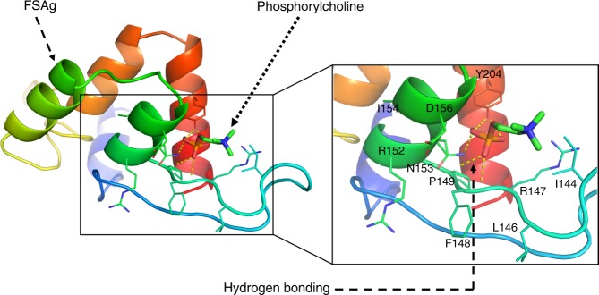
Fig. 7.
Molecular docking showing the interaction of microfilarial sheath antigen with phosphorylcholine. Crystal structure of phosphorylcholine was obtained from PubChem repository (PubChem ID: 135437). The 3D structure of microfilarial sheath antigen (FSAg) was obtained from our previously modelled structure13. The docking experiment was executed in silico employing Autodock Vina. Hydrogen bonds are depicted as yellow dashed lines. The conformation presented here possesses the negative binding energy (−4.0 kcal/mol)

