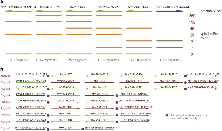
Figure 2.
TSD discovers the HBV integration events in the PLC/PRF/5 cells. (a) A genomic region identified by TSD with HBV integration event, which consists of 6 DNA fragments. The first line indicates the assembled HBV integrated region and the other lines indicate PacBio reads derived from this region. Each row can represent multiple PacBio reads if they have the same fragments composition; the number (right side) counts PacBio reads. The line colors and arrows indicate the mapped strands: darkgreen: forward; brown: reverse stand. (b) The HBV integration events discovered by TSD using PacBio sequencing data. TSD identified 9 HBV integrated regions, including multiple regions with complex HBV rearrangements. Most of the integration sites are validated by NGS study.

