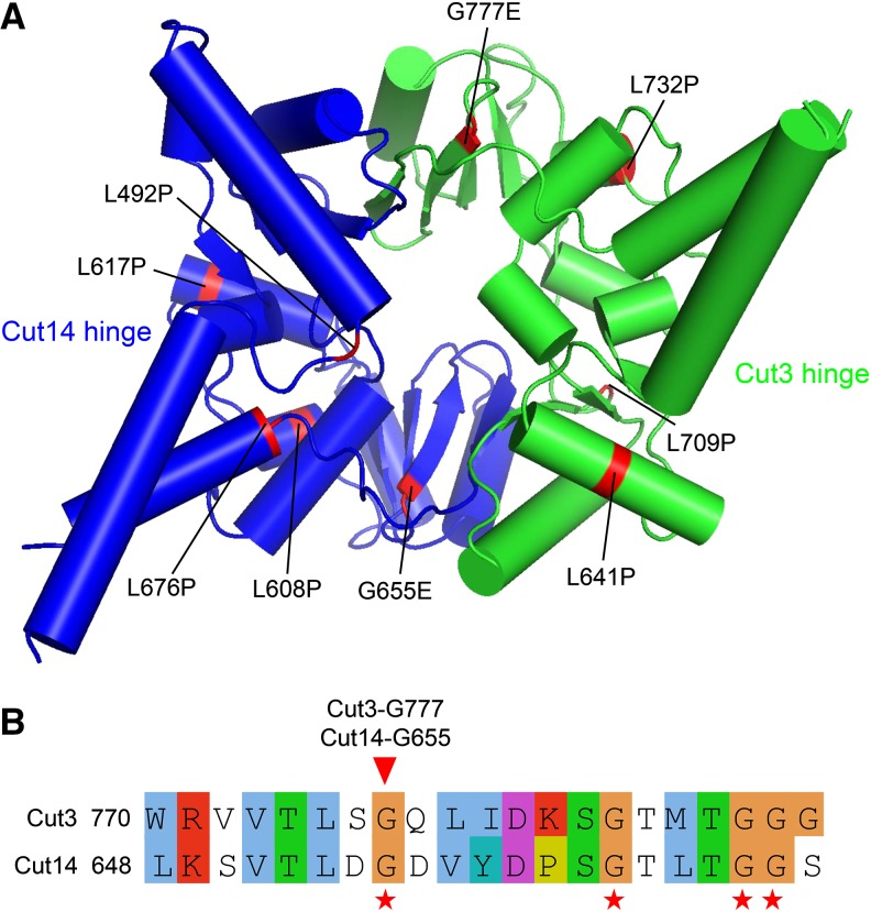
Figure 5.
Localization of newly isolated condensin hinge ts mutations in the 3D structure. (A) Mutations of the newly isolated condensin hinge ts mutants were mapped onto the atomic structure of the condensin hinge. The Cut14 hinge is in blue and the Cut3 hinge is in green. The broadly distributed mutations are highlighted in red. (B) Alignment of the Cut3 and Cut14 hinges around the conserved arrangement of glycine residues (GX6GX3GG sequence motif), which is normally found in hinge dimerization interfaces. Conserved glycines are marked with red asterisks below the alignment. Localization of Cut14-G655 and Cut3-G777 is indicated with the red arrowhead. Cut14-G655 and Cut3-G777 are in same position, but at different interfaces.

