. 2019 Feb 26;8(3):407–420. doi: 10.1039/c8tx00294k

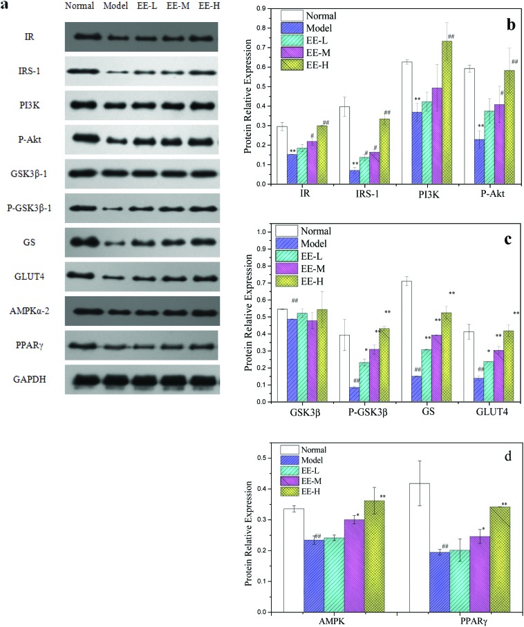
Fig. 9. Effects of EE on key protein expressions of liver insulin signalling pathway of mice: (a) western blot analysis of IR, IRS, PI3K, p-Akt, p-GSK3β, GSK3β, GS and glucose transporter 4 (GLUT4) protein expressions; (b) quantitative analysis of IR, IRS, PI3K and p-Akt protein expressions; (c) quantitative analysis of p-GSK3β, GSK3β, GS and GLUT4 protein expressions; (d) quantitative analysis of adenosine 5′-monophosphate-activated protein kinase (AMPKα-2) and peroxisome proliferator-activated receptor (PPARγ) protein expressions. Normal: normal group; Model: diabetic model group; EE-L, EE-M and EE-H: 150 mg kg–1, 250 mg kg–1 and 350 mg kg–1 EE-treated diabetic groups, respectively. *P < 0.05 and **P < 0.01 vs. normal group; #P < 0.05 and ##P < 0.01 vs. model group.

Fig. 9