. 2019 Mar 12;8(3):459–470. doi: 10.1039/c8tx00346g

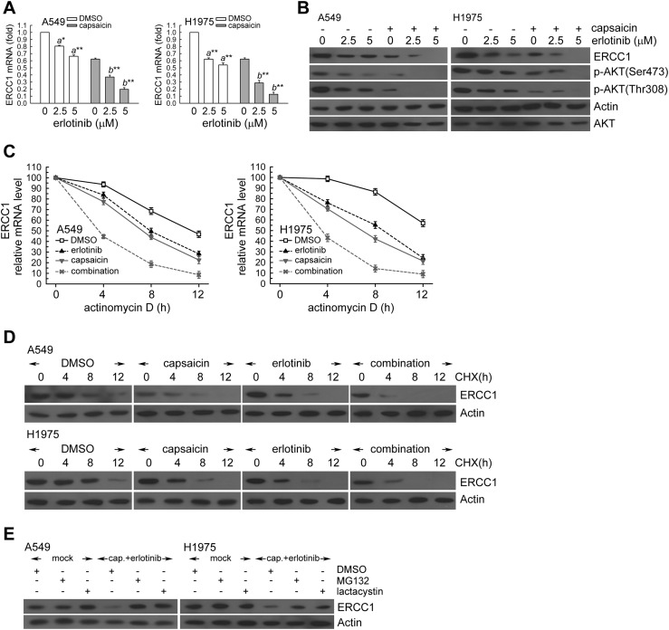
Fig. 6. Capsaicin decreased the ERCC1 protein and mRNA levels in erlotinib-exposed NSCLC cells. (A) A549 or H1975 cells (106) were cultured in complete medium for 18 h and then were exposed to capsaicin (25 μM) and erlotinib (2.5, 5 μM) for 24 h. After treatment, total RNA was isolated and subjected to real-time PCR for ERCC1 mRNA expression. The means ± standard deviation (SD) from four independent experiments. a** denotes p < 0.01, a* denotes p < 0.05 using Student's t-test for comparison between the cells treated with or without erlotinib. b** denotes p < 0.01, respectively, using Student's t-test for comparison between the cells treated with capsaicin alone or capsaicin and erlotinib combined. (B) After treatment as the above, cell extracts were examined by western blot for the determination of ERCC1, phospho-AKT(Ser473), actin, and AKT protein levels. (C) A549 or H1975 cells were treated with capsaicin (25 μM) and/or erlotinib (5 μM) for 12 h in the presence or absence of actinomycin D (2 μg mL–1) for 4, 8, or 12 h; total RNA was isolated and subjected to real-time PCR for ERCC1 mRNA expression. (D) The cells were exposed to capsaicin (25 μM) and/or erlotinib (5 μM) for 12 h followed by co-treatment with cycloheximide (CHX; 0.1 mg mL–1) for 4, 8, or 12 h. Whole-cell extracts were collected for western blot analysis. Capsaicin treatment triggers 26S proteasome-mediated proteolysis of ERCC1. (E) Capsaicin (25 μM) and erlotinib (5 μM) was added to A549 or H1975 cells for 18 h, then the cells were co-treated with 26S proteasome inhibitor MG132 (10 μM) or lactacystin (10 μM) for 6 h. Whole cell extracts were collected for western blot analysis.

Fig. 6