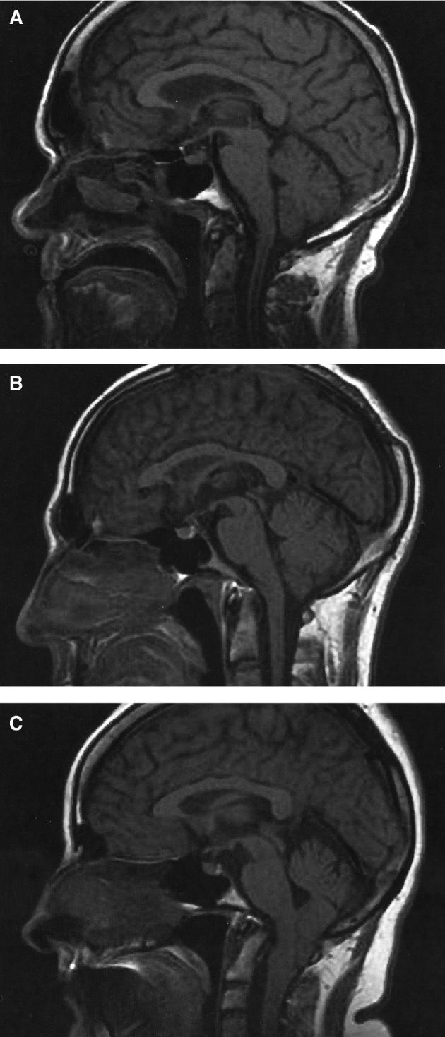
FIGURE 1.

Typical Chiari I (CM1), bCM1, and Chiari 0 malformation (CM0) on the sagittal cut MRI scans (from RT population). A, M70, CM0 with syringomyelia (the tonsils are at the level of the FM). B, F40, bCM1 with syringomyelia (tonsillar herniation = 3 mm). C, F41, CM1 without syringomyelia (tonsillar herniation = 17 mm).
