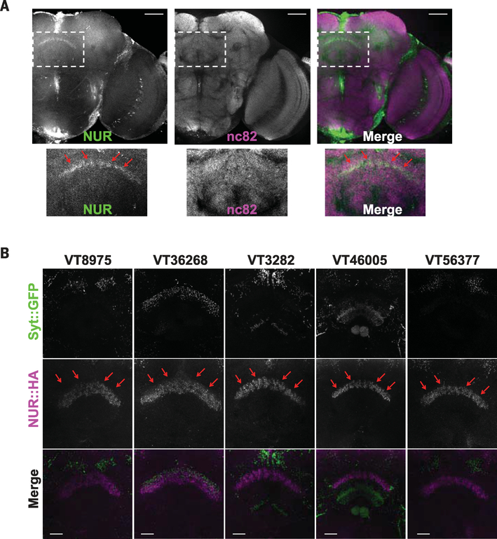
Fig. 6. NUR is induced by sleep loss and is localized to the dFSB area of the brain.
(A) Expression of endogenous NUR after sleep deprivation. Staining is for anti-NUR (green) and nc82 (magenta). The insets in the upper images are magnified below. Red arrows indicate expression of NUR in the dFSB. Scale bars, 50 μm. (B) Ectopic expression of NUR by different Gal4 drivers. Each Gal4 line was crossed with flies carrying UAS-syt::GFP and UAS-NUR::HA. Staining is with anti-GFP (top), anti-HA (middle), or merge (bottom). Note NUR expression in the dFSB area. Scale bars, 20 μm.

