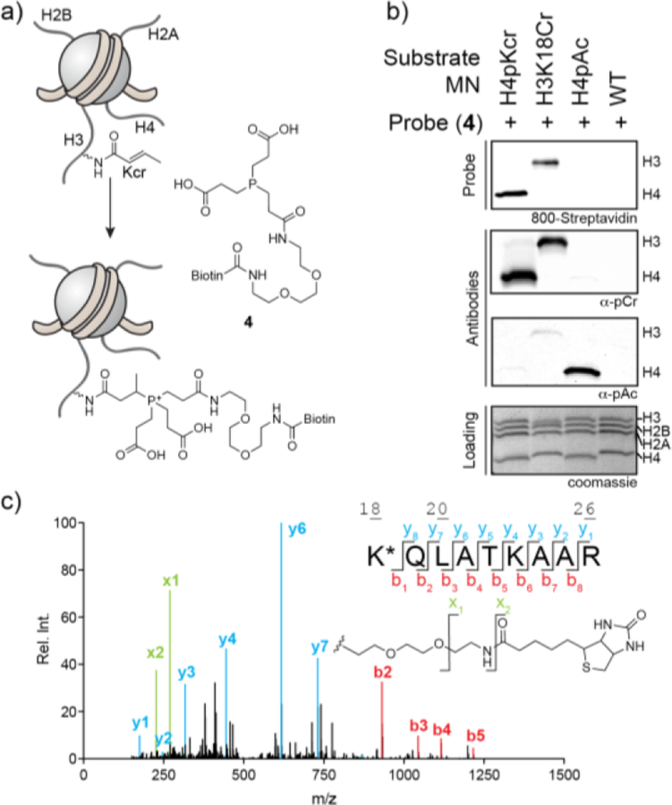
Figure 2.
A chemo-proteomic probe for protein lysine crotonylation. (a) Scheme depicting the application of TCEP analog 4 in the detection of protein crotonylation. (b) Semisynthetic mononucleosomes bearing indicated modifications were reacted with probe 4 (4 mM) before being analyzed by western-blot employing a biotin detection system (dye-conjugated streptavidin). For comparison, the same nucleosomes were analyzed by western blotting employing commonly used pan anti-lysine crotonyl (α-pCr) and pan anti-lysine acetyl (α-pAc) antibodies. A coomassie stained SDS-PAGE gel of the samples was used as a loading control. (c) Mass analysis of H3K18Cr conjugated with probe 4. Following the reaction, the protein was digested with ArgC and analyzed by LC-MS/MS. In addition to the expected y- and b-ions, fragmentation of the probe itself (lower right panel) is observed and denoted as x-ions.

