Fig. 3.
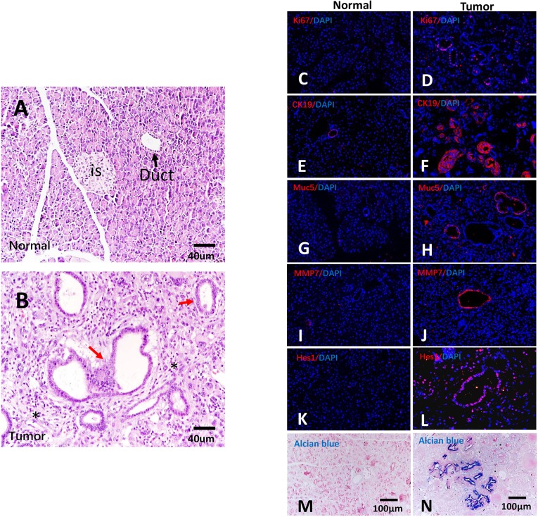
Characterization of pancreatic cancer in tree shrew. (A,B) H&E staining of normal (A) and cancerous pancreatic tissue (B) from tree shrew. is, islet; black arrow, normal duct; red arrows, glandular structures; asterisks, fibrous stroma. (C,D) Active cellular proliferation, as shown by Ki67 immunoreactivity (red). (E-L) Immunohistochemical detection of human pancreatic cancer markers CK19 (E,F), Muc5 (G,H), MMP7 (I,J) and Hes1 (K,L) (red). (M,N) Mucin proteins were stained by Alcian Blue. Normal tissue, C,E,G,I,K,M; tumor tissue, D,F,H,J,L,N.

