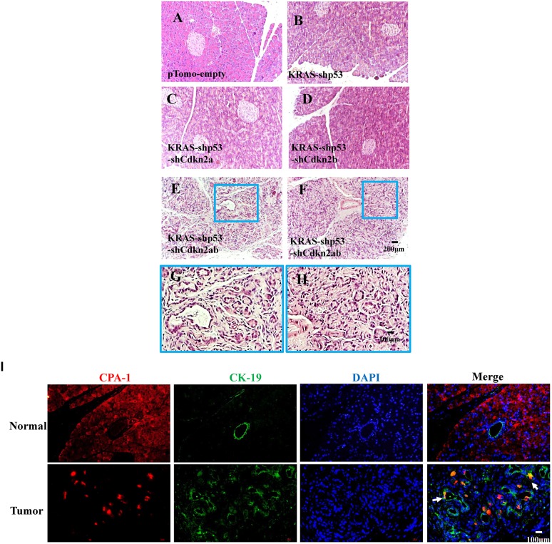
Fig. 4.
Detection of ADM in early stages of pancreatic cancer development in tree shrew. (A-H) Tissue was harvested 7 days after injection of the lentivirus, and H&E staining was performed in the pTomo-empty (A), KRAS-shTp53 (B), KRAS-shTp53-shCdkn2a (C), KRAS-shTp53-shCdkn2b (D) and KRAS-shTp53-shCdkn2a/b (E-H) groups. H&E staining shows an increase in the number of ducts in tissue infected by lentivirus KRAS-shTp53-shCdkn2a/b; the areas in the blue line boxes are shown enlarged below. (I) Immunohistochemical analysis of CPA1 (red) and CK19 (green) expression in normal tissue and tissue infected by lentivirus KRAS-shTp53-shCdkn2a/b; arrows indicate double-positive cells.

