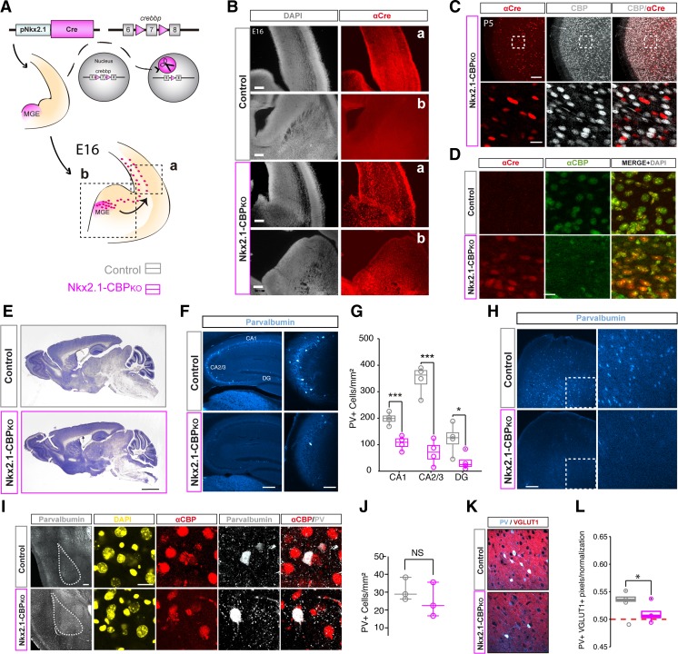
Fig. 1.
Nkx2.1-CBPKO mice have a reduced number of PV+ interneurons. A Scheme of genetic manipulation. The Nkx2-1 promoter drives the expression of Cre recombinase in MGE interneuron progenitors of Crebbpf/f mice, resulting in the elimination of exon 7 of Crebbp in these cells. B The expression of Cre-recombinase is detected in the expected areas in Nkx2.1-CBPKO E16 embryos Scale bars: 100 μm (a), 200 μm (b). C Double immunostaining for Cre recombinase and CBP in the parietal cortex of P5 mice. The detection of Cre recombinase coincides with the loss of CBP immunoreactivity. Scale bars: 100 μm (left), 20 μm (right). D Double immunostaining for Cre recombinase and CBP in striatal tissue and area that contains Nkx2-1-expressing cells in adult mice. Scale bar: 10 μm. E Representative images of Nissl staining in brain slices of Nkx2.1-CBPKO and control littermates. Scale bar: 1 mm. F Representative images of immunostaining with an anti-PV antibody. Scale bars: 200 μm (left) and 10 μm (right). G Quantification of the number of PV+ cells in the different hippocampal subfields. Nkx2.1-CBPKO mice have fewer PV+ cells in all substructures (DG, t6 = 2.55, p = 0.04; CA1, t6 = 5.17, p = 0.01; CA3, t6 = 7.63, p = 0.001). H Cortical areas, such as the frontal pole, also show a lower number of PV+ cells in Nkx2.1-CBPKO. Scale bar: 200 μm. I Immunostaining with an anti-PV antibody in the amygdala. Scale bars: 200 μm (left) and 20 μm (right). J The quantification of the number of PV+ cells in the amygdala revealed no significant difference. K Representative images of co-immunostaining with anti-PV and anti-VGlut1 antibodies. Scale bar: 50 μm. L Quantification of PV/VGLUT1 co-localization indicates that the glutamatergic innervation of PV+ interneurons is reduced in Nkx2.1-CBPKO (t6 = 2.62, p = 0.04, unpaired t test). *P < 0.05; ***P < 0.001

