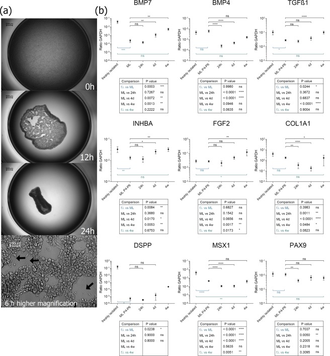
Figure 3.
(a) Kinetic of ultra-low attachment culture of DPCs. After 6 hours, long cell protrudings are formed between the cells (lowest picture, black arrows). (b) Analysis of gene transcription level of chosen marker genes during the condensation process. Depicted are relative expression values of BMP7, FGF2, PAX9, INHBA, TGFß1 and COL1A1 of the DPC condensates at 24 hours (24 h), 4 days (4d) and 4 weeks (4w) after induction of condensation culture. For comparison also expression values of freshly isolated DPCs (f.i.) and monolayer DPCs in different passages (ML P2-P6) are presented. ns - not significant; *P < 0.05; **P < 0.01; ***P < 0.001; ****P < 0.0001. N ≥ 5 biological replicates.

