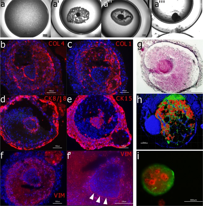
Figure 6.
Interaction of inductive condensates with epithelial cells (a): Human gingival keratinocytes do not condensate in vitro in ultra-ow attachment plates when cultured alone. In co-culture with 24 h-old DPC condensates they accumulate around the DPC condensate (a’), enwrap it (a”) and after 4 weeks a dense co-culture aggregate has formed (a”’). Immunohistological analyses (b–f’) show a structured organization within the co-culture aggregate after 4 weeks. Collagen Type IV (b) and Collagen Type I (c) are located in the middle cell layer with a visible accumulation on the tissue borders. Epithelial-specific keratins such as Cytokeratin 8/18 (d) and Cytokeratin 15 (e) are located in the outer layer of the co-culture aggregate and in the inner core. The mesenchyme marker Vimentin (f) is expressed by the middle layer of cells. Note the columnar arrangement of the epithelial cells in the inner core on the epithelium-mesenchyme border (f’, arrow tips). Hematoxylin-Eosin staining (g) shows an epithelial-band-like structure indicating the migration of the keratinocyte into the mesenchymal condensate. In (h) a co-culture aggregate was analyzed in 2-photon microscopy without labelling and an excitation wavelength of λ = 1300 nm using the SHG effect (red channel) and autofluorescence (blue and green channel). In (i) the two interacting cell types were labelled and tracked under fluorescent microscopy. DPCs were transduced to express GFP (green) and gingival keratinocytes were labelled with CellTrackerRed (red). Also, here the self-assembly and tissue organization in the in vitro culture is evident.

