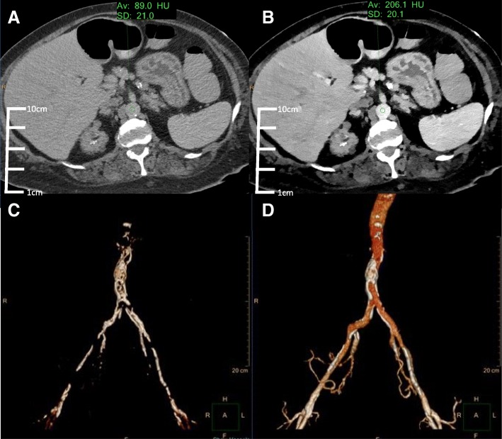
Fig. 1.
Conventional (a) and VMI 40 keV (b) axial SDCT images of case 1 TAVR planning. The 40 keV VMI data demonstrates increased aortic vascular contrast enhancement (HU: 206) compared to conventional CT (HU: 89). 3D volume renderings of the abdominal aorta from the conventional (c) and 40 keV VMI (d) data created using the same segmentation tools and workflow show better continuity and inclusion of the vascular lumen on 40 keV VMI compared to conventional CT

