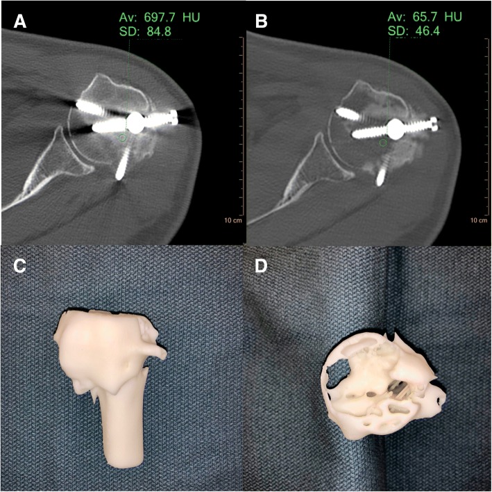
Fig. 5.
Conventional (a) and VMI 120 keV (b) axial SDCT images of case 4 left upper extremity trauma. There is significant reduction of metal artifact on the 120 keV VMI allowing for direct visualization of the metal hardware and associated incompletely healed fracture. c, d Various views of the 3D printed humeral head at 75% scale size using the Formlabs Form 2 standard white resin material. The cross-sectional view through the humeral head (d) demonstrates the fracture lines and track from the orthopedic intramedullary hardware

