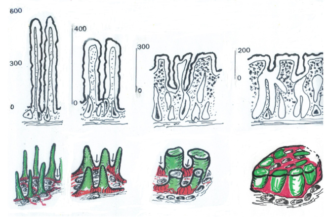
Figure 1.

These diagrams represent schematised accounts of the mucosal progression to “flattening”. The upper panel (a) gives some realistic indication of the reductions in mucosal height across the spectrum, from normal villi to the final mosaic platform.
The lower panel (b) provides a coloured description of the gradual upward growth of the inter-villous ridges (red) and their relationship to the villi (green) which are progressively shortened, by ~65–70%. Ultimately, the mosaic comprises a mixture of hypertrophied ridges which amalgamate the adjacent reduced villi: this seems to be the only intelligible way of explaining how mosaic plateaux are formed. Mosaic platforms, although containing villous cells, do not comprise individual villi, and cross sections of these plateaux must not be misinterpreted as such. The differential heights (a) give the proper interpretative clue.
