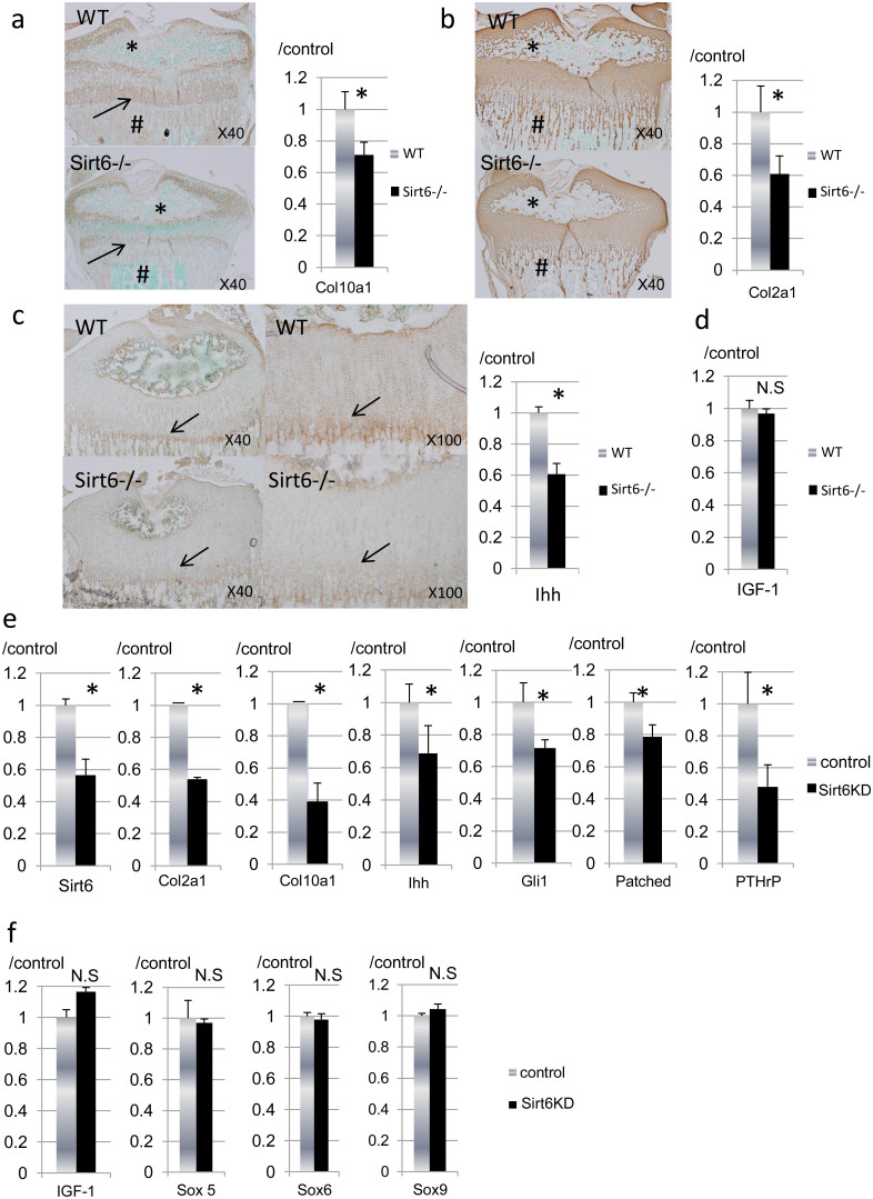
Figure 4. Effects of Sirt6 deficiency on chondrocyte differentiation.
(a–c) Immunohistological findings and real-time RT-PCR analysis for type X collagen, type II collagen and Ihh. Data are normalized to expression levels in WT cartilage and β-actin (n = 4). Note markedly reduced expression of type X collagen and type II collagen in the secondary ossification center (a, b, *) and primary spongiosa (a, b, #). Ihh expression was decreased at the prehyper-hypertrophic zone (arrows) in Sirt6−/− mice. mRNA expression of Col10a1 (a), Col2a1 (b) and Ihh (c) was significantly reduced in primary epiphyseal chondrocytes from Sirt6−/− mice. (d) mRNA expression of IGF-1 in primary epiphyseal chondrocytes was comparable between WT and Sirt6−/− mice. (e), (f) Primary epiphyseal chondrocytes were treated with Sirt6 siRNA. Total RNA was isolated from the cells and used in real-time RT-PCR with the indicated primers. When Sirt6 was knocked down by siRNA in primary chondrocytes, expression of Col2a1, Col10a1, Ihh, PTHrP, Gli1 and Patched were all decreased (e). (f) The expression of Sox5, Sox6 and Sox9 and IGF-1 was not affected by Sirt6 knockdown. The graph shows relative levels of gene expression. Values represent the mean ± SD of 3 samples per group. *; p < 0.05. N.S; no significant. Magnification is indicated in the figure.

