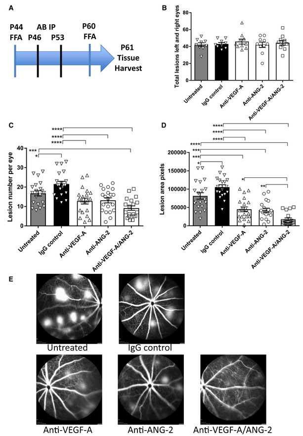
Figure 1. Reduction of vessel leakiness and lesion number by combined neutralization of VEGF‐A and ANG‐2 in the JR5558 mice using FFA .

(A) Study schematic. Baseline FFA was carried out at P44‐45. Antibody injections were given IP on P46 and P53. Post‐treatment FFA was at P60 and tissue harvest at P61. (B) Bar/scatter graph of baseline lesion numbers. Lesions were counted in all mice prior to study start. Combined total lesions from left and right eyes were calculated, and then, animals were assigned to treatment groups ensuring no statistically significant differences between groups (P > 0.05). (C, D) Bar/scatter graphs showing numbers of spontaneously occurring lesions (C) and area by fluorescence angiography (D) after two weekly doses of antibody (IgG, anti‐VEGF‐A, anti‐ANG‐2 at 5 mg/kg IP, and anti‐VEGF‐A/ANG‐2 at 10 mg/kg IP) followed by analysis a week after the last treatment. (E) Representative examples of fluorescence fundus angiograms from the untreated (top left), IgG control (top middle; 10 mg/kg), anti‐VEGF‐A (bottom left; 5 mg/kg), anti‐ANG‐2 (bottom middle; 5 mg/kg), and anti‐VEGF‐A/ANG‐2 (bottom right; 10 mg/kg) groups. Data information: SEM is shown as error bars with n = 9–10 animals (B) or n = 19–20 (C, D) eyes per group and significance indicated by asterisks using ANOVA (B: P > 0.05; C: P < 0.0001; D: P < 0.0001, followed by Newman–Keul's multiple comparison test in C, D). In (C), untreated is significantly different versus IgG control (*P < 0.05) and anti‐VEGF‐A/ANG‐2 (***P < 0.001). IgG control is significantly different versus anti‐VEGF‐A (****P < 0.0001), anti‐ANG‐2 (****P < 0.0001), and anti‐VEGF‐A/ANG‐2 (****P < 0.0001). In (D), untreated is significantly different versus IgG control (*P < 0.05), anti‐VEGF‐A (***P < 0.001), anti‐ANG‐2 (***P < 0.001), and anti‐VEGF‐A/ANG‐2 (****P < 0.0001). IgG control is significantly different versus anti‐VEGF‐A (****P < 0.0001), anti‐ANG‐2 (****P < 0.0001), and anti‐VEGF‐A/ANG‐2 (****P < 0.0001). Anti‐VEGF‐A/ANG‐2 is significantly different versus anti‐VEGF‐A (*P < 0.05) and anti‐ANG‐2 (**P < 0.01). FFA, fluorescein fundus angiography; P, post‐natal day; AB, antibody; IP, intraperitoneal; SEM, standard error of the mean; ANOVA, analysis of variance.Source data are available online for this figure.
