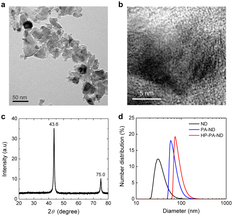
Figure 1. Characterization of 35-nm NDs and their bioconjugates.
(a) TEM image of the ND sample after air oxidation and strong oxidative acid treatment. (b) High-resolution TEM image of the ND particle, showing monocrystalline structure of the diamond matrix. (c) X-ray diffraction pattern of the ND powders exhibiting two distinct peaks at 2θ = 43.6° and 75.0°, corresponding to the (111) and (220) planes of crystalline diamond, respectively. (d) Size distributions of the NDs before and after surface modification with PA and subsequently with HP, measured by dynamic light scattering. The number-averaged diameters of NDs, PA-NDs, and HP-PA-NDs in water are 38 ± 4, 77 ± 2, and 91 ± 4 nm, respectively.

