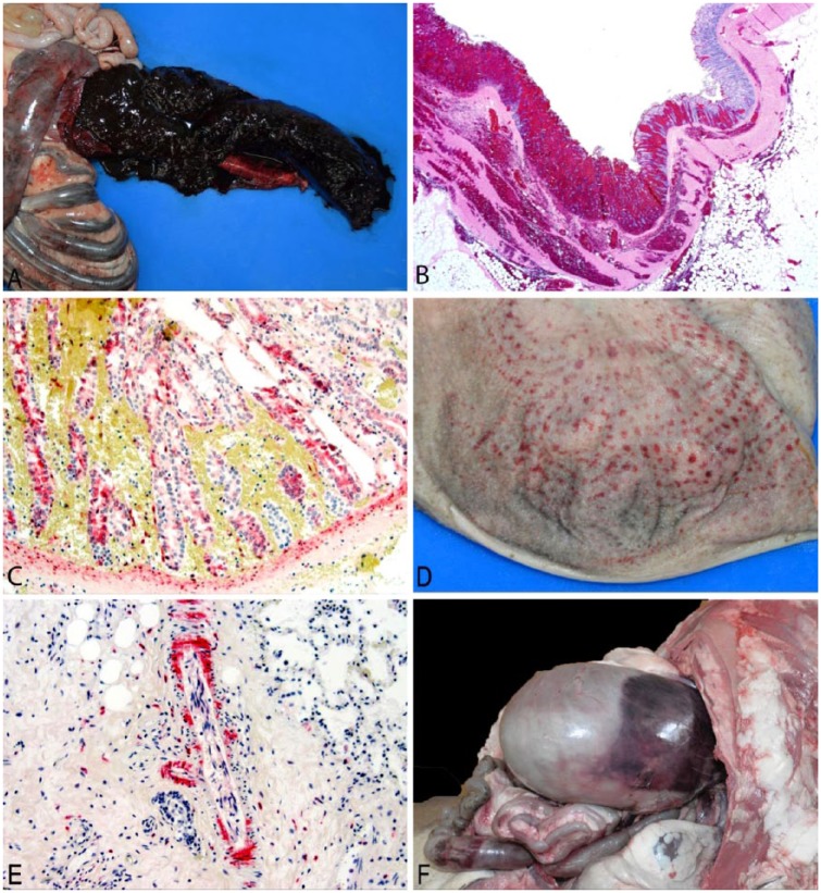
Figure 1.
Autopsy and histopathology from captive bighorn sheep with bovine viral diarrhea. A. Cecum opened to show necrohemorrhagic typhlitis, case 4. B. Histopathology of colon demonstrating hemorrhagic typhlocolitis from case 4 with full-thickness hemorrhage and mucosal necrosis. H&E. 20×. C. Immunohistochemistry (IHC) of panel B; positive staining for bovine viral diarrhea virus (BVDV) in mucosal epithelium and submucosal fibroblasts. BVDV IHC. 200×. D. Rumen opened to show necrohemorrhagic rumenitis, case 5. E. Immunohistochemistry of lung from case 6 showing diffuse staining of perivascular smooth muscle and fibroblasts, as well as a few scattered lymphocytes and macrophages. BVDV IHC. 200×. F. Suffusive hemorrhage on ruminal serosa, case 2.

