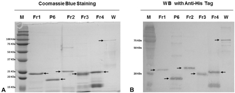
Figure 2.
Purification of African swine fever virus p72 polypeptide fragments. A. Sodium dodecyl sulfate–polyacrylamide gel electrophoresis (SDS-PAGE) with Coomassie blue staining. B. Western blot (WB) staining with anti-histidine monoclonal antibody. M = protein ladder; Fr1–Fr4 = polypeptide fragments; W = whole protein. Arrows indicate band showing the predicted size.

