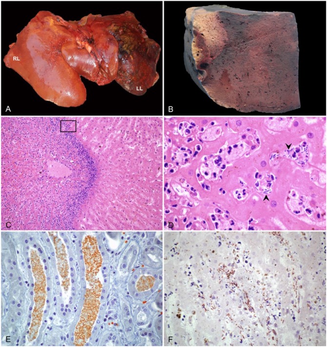
Figure 1.
Liver of a horse with infectious necrotic hepatitis. A. The parenchyma is dark-red to black with abundant fibrin covering the capsular surface of the left lobe (LL). RL = right lobe. B. On cut section of the left lobe, there is a large, clearly demarcated pale area of necrosis. C. Large random focus of coagulative necrosis, surrounded by a rim of neutrophils. H&E. D. Higher magnification of the area within the box in panel C, showing viable and degenerate neutrophils and large rods (arrowheads) among degenerate and necrotic hepatocytes. H&E. E. Orange-brown granules stained with Okajima stain for hemoglobin, within renal tubules. F. Clostridium novyi indirect immunoperoxidase staining in an area of hepatic necrosis, with numerous positive rods within the affected tissue.

