Figures 1–7.
Pulmonary adiaspiromycosis in a wild European rabbit (Oryctolagus cuniculus).
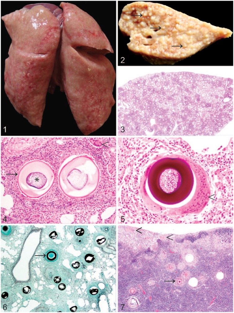
Figure 1. The lungs were firm and expanded by myriad granulomas.
Figure 2. The cut surface of the left caudal lung lobe was expanded by variably sized, coalescent, granulomas (arrows). Formalin-fixed tissue.
Figure 3. Numerous densely cellular foci surrounding spore-like adiaspores are present throughout the lung. H&E.
Figure 4. Adiaspores exhibit a bi- to trilaminar wall (arrow) comprising a thin, brightly eosinophilic outer layer, and a thick, pale eosinophilic inner layer surrounding a core of basophilic granular-to-foamy material (asterisk). Adiaspores are surrounded by heterophils, macrophages, multinucleate giant cells (arrowhead), lymphocytes, and plasma cells, in variable proportions, with interspersed fibroblasts. H&E.
Figure 5. The adiaspore wall is stained intensely positive with periodic acid–Schiff stain. Multifocally, adiaspores are surrounded by multinucleate giant cells with >40 nuclei (arrowhead).
Figure 6. The adiaspore wall exhibits intense staining with Grocott methenamine silver stain (arrow).
Figure 7. Similar adiaspores (arrow) are present within the tracheobronchial lymph node. Large multinucleate giant cells are also evident multifocally in this case within the subcapsular sinus (arrowheads).

