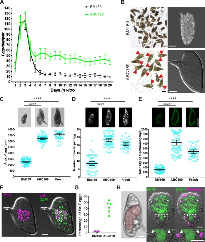
Fig 2. Eggs laid in ABC169 develop and hatch.
(A) Rate of egg production per worm pair in BM169 or medium ABC169 during in vitro culture. Dotted line indicates the point at which parasites begin laying morphologically abnormal eggs. n = 24 worm pairs in BM169 and n = 19 in ABC169, examined in 4 experiments. Error bars represent 95% confidence intervals. (B) Morphology of eggs (left, bright field; right, DIC) laid by paired adult females in BM169 or ABC169 on D20. Red arrows show early embryos in eggs laid in ABC169. (C–E) Quantification of the (C) size, (D) number of DAPI-labeled nuclei, and (E) autofluorescence intensity from eggs laid by freshly perfused female worms (Fresh, n = 41) and parasites cultured in BM169 (n = 61) or ABC169 (n = 59) on D20. In each group, eggs were harvested from 9 worm pairs across 3 separate experiments. ****p < 0.0001, t test. Error bars represent 95% confidence intervals. (F) EdU-labeled embryonic cells of an egg laid by a paired adult female in ABC169 on D20. (G) Percentage of eggs with clusters of cycling EdU+ embryonic cells from eggs laid between D16 and D22 by paired adult females in BM169 or ABC169. >2,000 eggs from 6 experiments. Error bars represent 95% confidence interval. (H) Miracidia from eggs laid in ABC169. Left, miracidium (pseudocolored red) inside an egg laid on D20 of culture in ABC169. Right, hatched miracidium from D20 egg labeled with DAPI and phalloidin. These miracidia appear grossly normal in morphology possessing 2 pairs of flame cells (arrows). Underlying primary data for panels A, C–E, and G can be found in S1 Data. Scale bars: B, F, H, 20 μm; C–E, 50 μm. ABC169, Ascorbic Acid, Blood Cells, Cholesterol, and BM169; BM169, Basch’s medium 169; D, day; DIC, differential interference contrast; EdU, 5-ethynyl-2′-deoxyuridine.

