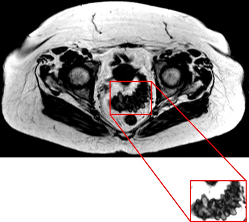
Fig 2. Fat only mDIXON image of the abdominal region of a patient with diverticular disease.
The portion of the colon with the diverticula is highlighted in the boxed image. The image was collected using a T1 weighted, water suppressed, sequence where adipose tissue appears bright and allows for its quantification. The colon and other organs appear darkly coloured, providing a contrast for visualisation of the diverticula.

