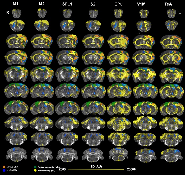
Fig 7. Mapping of ex vivo VBA-M and tract based connectivity maps.
To verify the overlap between morphometric changes detected ex vivo and circuits relevant to motor learning, we registered tract based connectivity maps [18] for individual seed regions to the minimum deformation template from our ex vivo study. We examined networks seeded in regions found to be enlarged in the contralateral (left) hemisphere, including the primary and secondary motor cortices (M1, and M2), as well as the primary and secondary somatosensory cortices (S1 –the forelimb region, S2), caudate putamen (CPu), V1M primary visual cortex (monocular); and the temporal association cortex (TeA). Significant clusters for in vivo morphometry at the terminal point are shown in blue, the interaction of time by group (trained versus not trained) in green, and the morphometric results from ex vivo specimens are shown in orange. These clusters showed good overlap with the tract density maps (TD).

