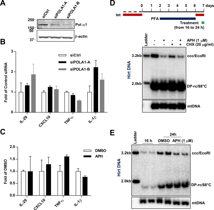
Fig 5. The effect of APH and CD437 on cccDNA formation is rapid and direct.
(A) Seventy-two hours after transient transfection of indicated siRNAs in HepAD38 cells, protein levels of Pol α1 and β-actin were determined by Western blot assays. (B) The mRNA levels of IL-29, CXCL10, TNF-α and IL-1β were measured by qRT-PCR and presented as fold over that in cells transfected by control siRNA (mean ± SD; n = 3). (C) HepAD38 cells were mock-treated (DMSO) or treated with 1 μM APH for 8 h. Expression of the indicated cytokine mRNA and β-actin mRNA were measured by qRT-PCR and were presented as fold over that in DMSO treated cells (mean ± SD; n = 3). (D and E) HepAD38 were cultured in the presence of 2 mM PFA from day 2 to day 6 after tet removal. From day 6, cccDNA synthesis was initiated by removing PFA for 24 h, with indicated compound treatments during the last 8 h (16 to 24 h) of cccDNA synthesis. Hirt DNA was extracted and resolved by Southern blot hybridization, with mtDNA as a loading control.

