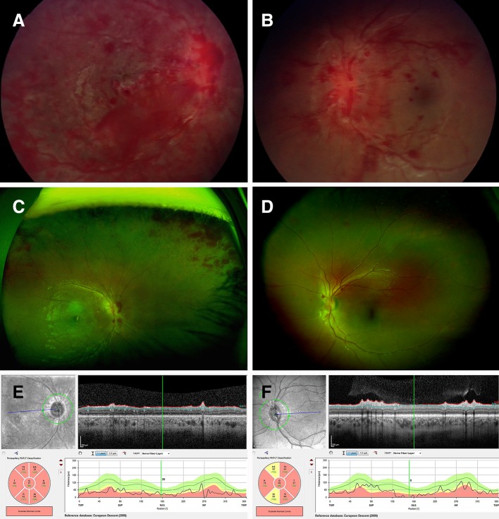
Figure 1.
Ocular images of child with ADEM. (A,B) Retinal images on presentation right (A) and left (B), demonstrating extensive retinal haemorrhages involving multiple layers of the retina, right eye greater than left, dilated and tortuous retinal venules, and optic nerve head swelling left eye, but not visible in the right eye. (C,D) Optos ultra-widefield retinal photos day 10 postpresentation, showing improvement, but persistent haemorrhages particularly peripapillary and in the peripheral retina, with optic nerve head swelling and cotton wool spots. (E,F) Retinal nerve fibre layer 18 months’ postpresentation demonstrating significant thinning both eyes, right >left. ADEM, acute disseminated encephalomyelitis.

