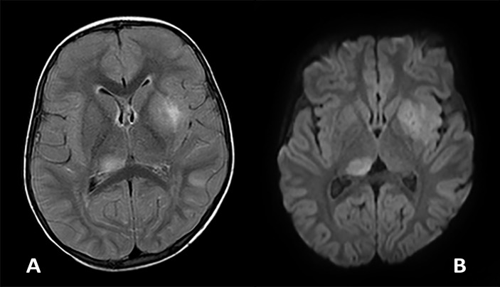
Figure 2.
Axial FLAIR (A) and diffusion (B) images demonstrating two representative lesions within the right thalamus and left lentiform nucleus and external capsule at day 2 of presentation. The lesions are asymmetrical, ill-defined and T2 hyperintense on the FLAIR and have increased diffusivity (bright signal) on the diffusion. FLAIR, fluid-attenuated inversion recovery.

