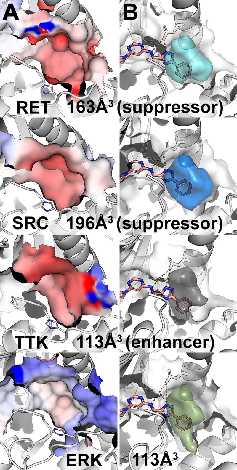
Fig 3. Visualization of DFG-pockets.

(A) Electrostatic potential (red, negative potential; blue, positive potential) on the surface of the DFG-pocket in various kinases, including the suppressors RET and SRC, the enhancer TTK, and ERK. (B) Accessible volume of the DFG-pocket (colored volume) for potential type-II kinase inhibitor. Hit molecule 1 is depicted in pink sticks. Broken yellow lines indicate hydrogen bonds.
