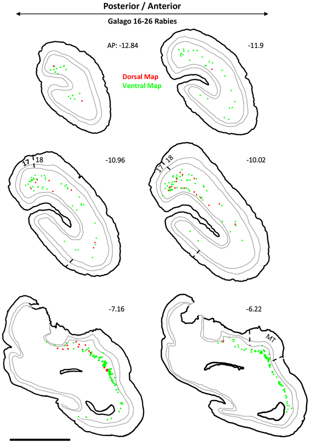
Figure 8:
Neurons labeled in visual cortex after rabies virus injections into the dorsal and ventral pulvinar maps in galago 16–26. The locations of labeled cells projecting to the dorsal map are shown in red while those projecting to the ventral map are shown in green. The coronal sections are arranged in a posterior to anterior sequence. The internal borders drawn in grey are the layer 4/5 and 5/6 borders. Areas 17, 18, and MT are delineated on appropriate sections. AP level indicated for each section.

