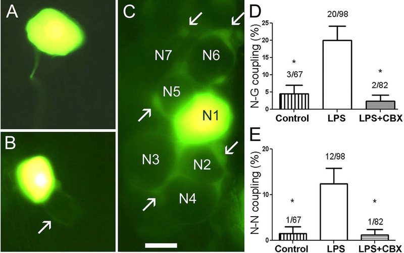
Figure 1. Lucifer yellow (LY) injection into neurons in intact TG from LPS-treated mice reveals homocellular and heterocellular gap junction-mediated coupling.
A. Injected neuron in TG isolated from control animal is typically not dye-coupled to any cell. B. In a very small number of cases an injected neuron in a control TG was found to be coupled to a surrounding SGC and to the SGC sheath surrounding a neighboring neuron (arrow indicates the SGC envelope coupled to the injected neuron). C. TG from a mouse that had been injected with LPS (2.5 mg/kg) eight days earlier. This is an example of the larger proportion of cases in which N-G coupling was present. The injected neuron (N1) was dye-coupled to SGCs around at least six other neurons (N2–N7). Some of the coupled SGCs are indicated by arrows. Calibration, 20 μm. D. Summary of frequency of observing LY dye transfer to SGC (G) when a neuron was injected. In control conditions such coupling was rare (about 5%), but after LPS injection, N-G coupling increased four-fold. The addition of CBX (100 μM) to the medium during LY injection into cells in ganglia from LPS injected mice reduced coupling incidence to about 3% (* p<0.01 between control and LPS treatment and between LPS treatment and LPS + CBX). E. Effects of LPS injection on coupling between neurons in intact TG. In controls the prevalence of such coupling was low (<2%); however, at eight days after LPS injection we observed neuron-neuron coupling in about 13% of the cases. The gap junction blocker CBX blocked the intercellular spread of LY, confirming that gap junctions were involved. (* p<0.05: Fisher’s exact test).

