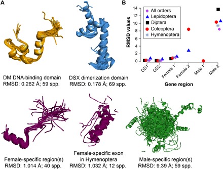
Fig. 5. Protein structural conservation in various domain and sex-specific regions of dsx in insects.

Drosophila melanogaster crystal structures for OD1 and OD2 were used as a reference for protein modeling from the dsx sequences. (A) Superimposition of protein structures in each exonic region. RMSD as a measure of structural deviation between protein backbones and the number of species used for protein modeling are also shown. (B) Comparison of protein structural differences across exonic regions and insect orders, which highlights male-biased evolution of the Dsx protein. Since protein structures are relatively similar in sister species, only representatives of each genus were chosen for this comparison (see table S1 for the list of species used for modeling and fig. S1 for structures of domains and sex-specific regions of each insect order).
