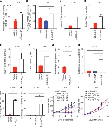
Fig. 5. hsBCL9CT-24 reactivates anticancer immunity and overcomes resistance to PD-1 inhibitors.

(A) BALB/c mice inoculated with CT26 tumors were treated with vehicle control or hsBCL9CT-24 (20 mg/kg) via i.p. injection, QD over 14 days (n = 4 per cohort). The percentage of CD103+ cells among CD45+CD11c+ DC cells in tumor was analyzed (**P < 0.01). (B) Percentage of CD103+ cells among CD45+CD11c+ DC cells in wild-type (transduced without shRNA), NT shRNA, or β-cat shRNA transduced CT26 tumors (*P < 0.05, **P < 0.01). (C) qRT-PCR measurement of CCL4 expression in CT26 cells treated with vehicle or hsBCL9CT-24 at 5 μM for 24 hours (**P < 0.01). (D) qRT-PCR measurement of CCL4 expression in CT26 cells transduced with NT shRNA or β-cat shRNA (**P < 0.01). (E) Fluorescence-activated cell sorting analysis of the ratio of CD8+ to CD45+ T cells in the tumors described in (A). (F) Ratio of CD8+ cytotoxic T cells over Treg cells in the tumors described in (A) (**P < 0.01). (G) Fold change of granzyme B+CD8+ T cells among the overall CD8+ T cell population before and after hsBCL9CT-24 treatment (**P < 0.01). (H) Fold change of granzyme B+CD8+ T cells among overall CD8+ T cell population in CT26 WT, NT shRNA, and β-cat shRNA transduced tumors (**P < 0.01). (I) Fold change of CD8+CD44+CD62L− cells (effector CD8+ cells) among the overall CD8+ T cell population before and after hsBCL9CT-24 treatment (**P < 0.01). (J) Fold change of effector CD8+ cells among the overall CD8+ T cell population in CT26 WT, NT shRNA, and β-cat shRNA transduced tumors (**P < 0.01). Statistical significance of differences between groups was determined by unpaired Student’s t test. (K) Combination treatment of hsBCL9CT-24 and anti–PD-1 Ab resulted in almost complete regression in the LLC1 model. C57BL/6 mice were inoculated with LLC1 cells via single flank implantation and treated with immunoglobulin G (IgG), hsBCL9CT-24 (25 mg/kg, QD), anti–PD-1 Ab [twice weekly (BIW)], and hsBCL9CT-24 + anti–PD-1 Ab as indicated after tumor volume reached 30 mm3 (n = 4 per cohort) (**P < 0.01). (L) Combination treatment of hsBCL9CT-24 and anti–PD-1 Ab resulted in significant tumor reduction in the 4T1 model. BALB/c mice were inoculated with 4T1 cells via mammary gland inoculation and treated with IgG, hsBCL9CT-24 (20 mg/kg) QD, anti–PD-1 Ab, BIW, and hsBCL9CT-24 + anti–PD-1 Ab as indicated after tumor volume reached 20 mm3 (n = 4 per cohort) (**P < 0.01). Statistical significance of differences in tumor growth assays was determined by two-way ANOVA. Results were denoted as means ± SEM for experiments performed in triplicate, and each experiment was repeated twice.
