Figure 2.

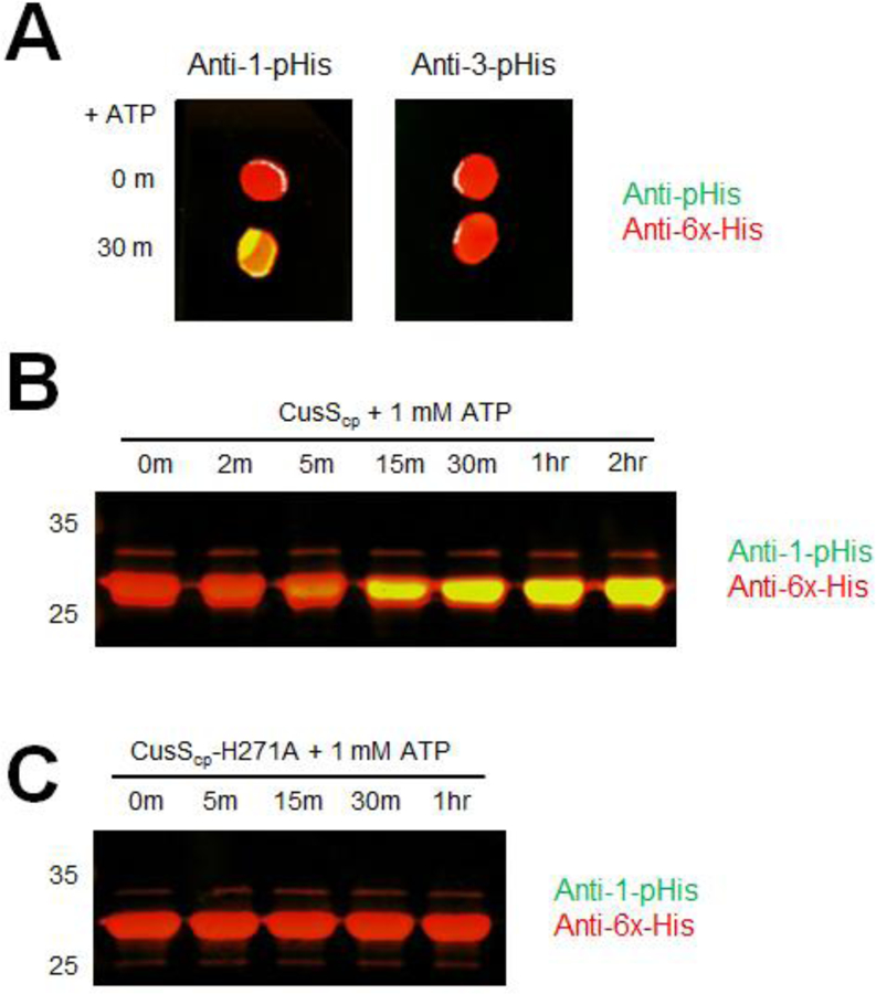
In vitro autophosphorylation of CusScp wild-type and CusScp-H271A. (A) Dot blot analysis of CusScp. CusScp was diluted to 10 μM in kinase buffer and autophosphorylation was initiated by adding ATP and incubated at 37°C. Samples were collected at 0 and 30 min, then 2 μL of each was spotted on PVDF membrane and probed with anti-1-pHis and anti-His tag antibodies simultaneously (left), or with anti-3-pHis and anti-His tag antibodies simultaneously (right). (B) CusScp (25 μM) was diluted in kinase buffer containing ATP to a final concentration of 1 mM, and reactions were incubated at 37°C. Samples were collected at the indicated times (2 min to 2 hr). (C) CusScp-H271A (25 μM) was diluted in kinase buffer containing ATP to a final concentration of 1 mM, and reactions were incubated at 37°C. Samples were collected at the indicated times (5 min to 1 hr). The reactions were terminated by adding 2X SDS sample buffer and were loaded onto SDS-PAGE. After electrophoresis, the proteins were transferred onto PVDF membranes and analyzed by Western blot probed with anti-1-pHis and anti-His-tag antibodies simultaneously. For all panels, each experiment was repeated at least three times; a representative experiment is shown.
