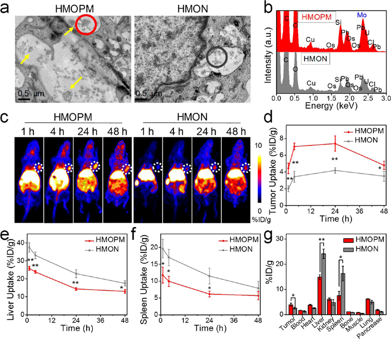
Figure 4.
Enhanced tumor accumulation based on the acidity-driven in situ self-assembly of HMOPM. (a) Bio-TEM images of U87MG cells incubated with HMOPM or HMON. Scale bar, 0.5 μm. Many HMOPM aggregates (marked with yellow arrows) were clearly observed in the endosomes at pH 5.0−6.0, while no apparent assemblies were found in the HMON-treated cells. (b) EDS elemental analysis of the correspondingly circled regions in (a). (c) Representative PET images of U87MG tumor-bearing mice at 1, 4, 24, 48 h postinjection (p.i.) of the 64Cu-labeled HMOPM or HMON. Tumors were circled with white dots. (d−f) PET quantification on tumor (d), liver (e), and spleen (f) uptake (n = 4, mean ± s.d.). (g) Biodistribution of the 64Cu-labeled HMOPM or HMON at 48 h p.i. (n = 4, mean ± s.d.). Significantly enhanced tumor accumulation and strikingly decreased liver and spleen uptake were observed in the HMOPM-treated mice comparing to the HMON-treated ones. * P < 0.05. ** P < 0.01.

