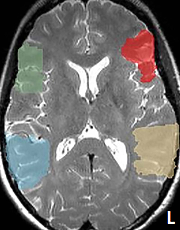
Figure 1.
One example patient’s language regions-of-interest overlaid onto the structural image. The language regions-of-interest (ROIs) defined in the Montreal Neurological Institute space were reversed-normalized to this patient’s own brain space. The anterior ROIs are shown in red for the left hemisphere and green for the right hemisphere. The posterior ROIs are shown in yellow for the left hemisphere and blue for the right hemisphere.

