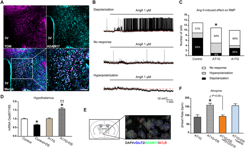
Figure 5. Ang-II-induced sympatho-excitation was reduced in mice with ADAM17 knockdown in glutamatergic neurons.
(A) Within the PVN, tdTomato-expressing glutamatergic neurons (magenta) show robust expression of ADAM17 (cyan), as indicated by the merged panel (white). The scale bar represents 38 μm. (B) Representative traces demonstrating depolarization, no response and hyperpolarization of PVN kidney-related glutamatergic neurons from control mice following Ang-II application. All recorded neurons were visualized through tdTomato and PRV-152 labeling (see Fig. S3). (C) Distribution of the various responses to Ang-II application on resting membrane potential of kidney-related glutamatergic PVN neurons for control (n=13 cells from 6 mice), AT1G (n=11 cells from 5 mice) and A17G (n=10 cells from 4 mice) groups. (D) Glutamate decarboxylase 1 (Gad67) gene expression (qRT-PCR) in hypothalamus of control and A17G mice (n=6 mice/group). (E) Fluorescent in situ hybridization (FISH) on a PVN brain section for visualization of ADAM17 (green) and AT2R (red) in glutamatergic neurons (vGluT2-labeled, blue); the nuclei are labeled with DAPI (grey). Magnification: 400X. (F) Cardiac vagal tone was studied through intraperitoneal injections of a muscarinic antagonist (atropine, 1 mg/kg) in AT1G mice (n=10 mice/group). Saline or PD123319 (5 μg/mouse) was given to the DOCA-salt-treated AT1G mice through intracerebroventricular injection, 20 min before atropine injection (n=6 mice/group). Statistical significance: Chi-square test: *P<0.05 vs. control; Two-way ANOVA: *P<0.05 vs. respective sham or baseline, ††P<0.01 vs. control+DS. Student’s t-test: AT1G+DS vs. AT1G+DS+PD123319, P<0.05. DS: DOCA-salt.

